Velocity and Shear Stress Measurements in the Lymphatic System
Recently, our group has developed an imaging
technique to measure lymph flow and sheer stress in a minimally invasive
system compared to previous methods.
The lymphatic system plays a crucial role in lipid transport and immune cell transportation, making this system one of the most important transport systems in the body. However, its functionality is not well understood yet. Since the lymphatic system’s main role is transport, the key parameter to better comprehension of its functionality lies in studying the flow in the lymphatic vessels. Many groups tried to study the lymphatic system by measuring the contractile activity or other parameters but the flow remains the only parameter directly related to the transport function of this system. Others tried to measure the flow by cannulating lymph vessels but this method is sturdily invasive and damages the lymphatic system, also it doesn’t give the real exact flow in the vessels. Our group has developed an imaging system to directly measure lymph velocity, which leads to flow, in rat mesenteric micro-lymphatic vessels. Although this technique is invasive, it does not damage the lymphatic vessels like cannulation methods. Plus, this method gives exact measurements of lymph velocity, flow, and sheer stress.
 |
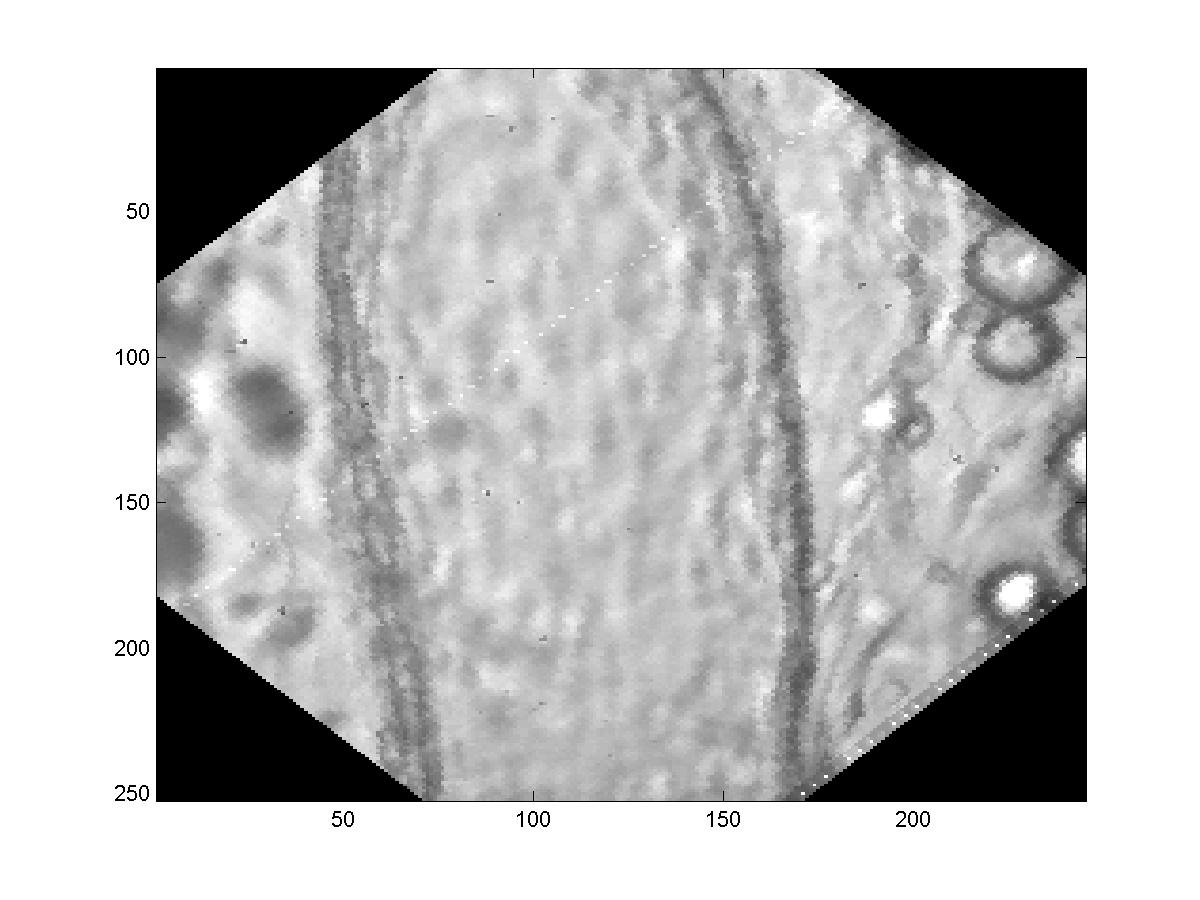 |
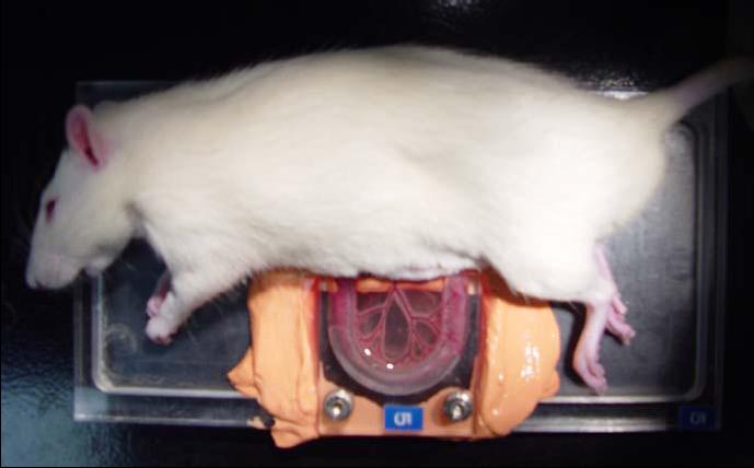
| Preparation of rat mesentery for high speed imaging (500 fps) | Image of lymphocytes traveling through mesenteric lymph vessel |
The technique is based on optical video microscopy. Images are
acquired using a high speed CCD camera and processed with a complex
algorithm that uses image correlation to estimate the displacement of
lymph particles. Our camera captures images at 500 fps (typical video
rate is 30 fps) which is a must due to the high velocity peaks that
occur during the systolic phase of the contraction cycle of the lymph
vessel. Several filtering techniques are then applied to the data to
eliminate motion artifacts and noise that is a big factor with in situ
experiments. We recently optimized our algorithm and got the processing
time of a typical data period down to 3 minutes that is considered near
real time in comparison to our previous versions.
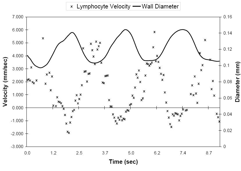 |
| Lymph velocity (*) and diameter (-) measurements are out of phase, showing the correlation with contraction of the lymph vessel and lymphocyte velocity. The negative velocity indicates backflow of the lymphocyte during diastole. |
In the future, this system will be used to monitor acute changes in
microlymphatics due to different imposed parameters. Also we plan on
expanding the functionality of this system to be able to monitor the
contractile activity and study its propagation and origination which is
still an underdeveloped area.
Note: All pictures were obtained from the reference below.
Collaborators:
Dr. Dave Zawieja and Dr. Anatoliy Gashev with the Department of Systems Biology and Translational Medicine, Texas A&M Health Science Center
Dr. James Moore, Biomedical Engineering, TAMU.
References:
Dixon, J. B. (2006). A Biomedical Engineering Approach to Investigating Flow and Wall Shear Stress in Contracting Lymphatics Biomedical Engineering. College Station, Texas A&M University. Ph.D.
For more information, contact Tony Akl
Department of Biomedical Engineering | Dwight Look College of Engineering | Texas A&M University